Wednesday, August 18, 2010

fOrum in cLass..


On 9th August 2010, our lecturer English do some activity for us. We are divided into seven group, whereby each group have their own topic to present. We are the first group that have been chosen to present the forum. our forum topic us about in effective way to curb the dengue cases in the school.
This is one of the speaking practice before Malaysian University English Test (MUET).
so that from the forum, we can practice how to speak with the proper way and correct grammar.

HeaD LicE

What are head lice?

Head lice are parasites that can be found on the heads of people. Infection with head lice is called pediculosis.
(The head louse, Pediculus humanus capitis, is different from the lice that cause body and pubic-hair infections.)
View Image

Who is at risk for getting head lice?

Anyone who comes in close contact with someone who already has head lice, or even their contaminated clothing and other belongings, is at risk for acquiring head lice. Preschool and elementary school children (3-10 years of age) and their families are infected most often. Girls contract head lice more often than boys; women more often than men. African-Americans rarely acquire head lice.

How in the world does a child get head lice?

A child can contract head lice in a number of ways:
  • contact with an already infested person. Personal contact is common during play, school, or sports activities, and at school, home, slumber parties, or camp;
  • wearing infested clothing, such as hats, scarves, coats, sports uniforms, or hair ribbons;
  • using infested combs, brushes, or towels; or
  • lying on a bed, couch, pillow, carpet, or stuffed animal that has recently been in contact with a person with lice.

What do head lice look like?

There are three forms of lice, namely the nit, the nymph, and the adult louse:
Nit: Nits are lice eggs. Nits are hard to see and are often confused with dandruff or hair spray droplets. Nits are found firmly attached to the hair shaft. They are oval and usually yellow to white. Nits take about a week to hatch.
Nymph: The nit hatches into a baby louse called a nymph. It looks like an adult head louse, but is smaller. Nymphs mature into adults about seven days after hatching. To live, the nymph must feed on blood.
Adult: The adult louse is about the size of a sesame seed, has six legs, and is tan to grayish-white in color. In people with dark hair, the adult louse looks darker. Females lay nits; they are usually larger than males. Adult lice can live up to 30 days on a person's head. To live, adult lice need to feed on blood. If the louse falls off a person, it dies within two days.
Go to fullsize image

Where are head lice most commonly found?

Head lice are most frequently located on the scalp behind the ears and near the neckline at the back of the neck. Head lice hold on to hair with hook-like claws that are found at the end of each of their six legs. Head lice are rarely found on the body, eyelashes, or eyebrows.

What are the signs and symptoms of head lice infestation?

The signs and symptoms are
  • a tickling feeling of something moving in the hair;
  • itching (caused by the an allergic reaction to the bites);
  • sores on the head (caused by scratching);
  • these sores on the head can sometimes become infected; and
  • irritability (a very nonspecific thing, to be sure).

Go to fullsize image

How is a head lice infestation (pediculosis) treated?

For effective elimination of head lice, the infested individual, family members that are also infested, and the home must all be treated.
Treatment of the individual and the infected family members:
Over-the-counter (OTC) or prescription medications are used to treat the affected people and their families. Follow these treatment steps:
  1. Remove all clothing.
  2. Apply lice medicine, also called pediculicide, according to the label instructions. If your child has extra long hair, you may need to use a second bottle. WARNING: Do not use a cream rinse or combination shampoo/conditioner before using lice medicine. Do not re-wash hair for one to two days after treatment.
  3. Have the infested person put on clean clothing after treatment.
  4. If some live lice are still found eight to 12 hours after treatment, but are moving more slowly than before, do not retreat. Comb dead and remaining live lice out of the hair. The medicine sometimes takes longer to kill the lice.
  5. If, eight to 12 hours after treatment, no dead lice are found and lice seem as active as before, the medicine may not be working. See your health-care provider for a different medication and follow their treatment instructions.
  6. Nit (head lice egg) combs, often found in lice medicine packages should be used to remove nits and lice from the hair shaft. Many flea combs made for cats and dogs are also effective.
  7. After the initial treatment, check, comb, and remove nits and lice from hair every two to three days.
  8. Re-treat in seven to 10 days.
  9. Check all treated people for two to three weeks until you are sure all lice and nits are gone.
Treating the house:
Treating the whole house is a laborious but important task. Follow these steps:
  1. Machine wash all washable clothing and bed linens that the infested person touched during the two days before treatment (to kill the lice and nits). Use the hot water cycle (130 degrees F; 55 degrees C) to wash clothes. Dry laundry using the hot cycle for at least 20 minutes.
  2. Dry clean clothing that is not washable (coats, hats, scarves, etc.), or store all clothing, stuffed animals, comforters, etc., that cannot be washed or dry cleaned into a plastic bag and seal it for two weeks.
  3. Soak combs and brushes for one hour in rubbing alcohol, Lysol, or wash with soap and hot (130 degrees F; 55 degrees C) water.
  4. Vacuum the floor and furniture. Do not use fumigant sprays. (They can be toxic if inhaled.)

GasTriTis

What is gastritis?

Gastritis is a condition in which the stomach lining—known as the mucosa—is inflamed. The stomach lining contains special cells that produce acid and enzymes, which help break down food for digestion, and mucus, which protects the stomach lining from acid. When the stomach lining is inflamed, it produces less acid, enzymes, and mucus.
Gastritis may be acute or chronic. Sudden, severe inflammation of the stomach lining is called acute gastritis. Inflammation that lasts for a long time is called chronic gastritis. If chronic gastritis is not treated, it may last for years or even a lifetime.
Erosive gastritis is a type of gastritis that often does not cause significant inflammation but can wear away the stomach lining. Erosive gastritis can cause bleeding, erosions, or ulcers. Erosive gastritis may be acute or chronic.
The relationship between gastritis and symptoms is not clear. The term gastritis refers specifically to abnormal inflammation in the stomach lining. People who have gastritis may experience pain or discomfort in the upper abdomen, but many people with gastritis do not have any symptoms.
The term gastritis is sometimes mistakenly used to describe any symptoms of pain or discomfort in the upper abdomen. Many diseases and disorders can cause these symptoms. Most people who have upper abdominal symptoms do not have gastritis.

View Image

What causes gastritis?

Helicobacter pylori (H. pylori) infection causes most cases of chronic nonerosive gastritis. H. pylori are bacteria that infect the stomach lining. H. pylori are primarily transmitted from person to person. In areas with poor sanitation, H. pylori may be transmitted through contaminated food or water.
In industrialized countries like the United States, 20 to 50 percent of the population may be infected with H. pylori.1 Rates ofH. pylori infection are higher in areas with poor sanitation and higher population density. Infection rates may be higher than 80 percent in some developing countries.1
The most common cause of erosive gastritis—acute and chronic—is prolonged use of nonsteroidal anti-inflammatory drugs (NSAIDs) such as aspirin and ibuprofen. Other agents that can cause erosive gastritis include alcohol, cocaine, and radiation.
Traumatic injuries, critical illness, severe burns, and major surgery can also cause acute erosive gastritis. This type of gastritis is called stress gastritis.
Less common causes of erosive and nonerosive gastritis include
  • autoimmune disorders in which the immune system attacks healthy cells in the stomach lining
  • some digestive diseases and disorders, such as Crohn’s disease and pernicious anemia
  • viruses, parasites, fungi, and bacteria other than H. pylori
View Image

What are the symptoms of gastritis?

Many people with gastritis do not have any symptoms, but some people experience symptoms such as
  • upper abdominal discomfort or pain
  • nausea
  • vomiting
These symptoms are also called dyspepsia.
Erosive gastritis may cause ulcers or erosions in the stomach lining that can bleed. Signs of bleeding in the stomach include
  • blood in vomit
  • black, tarry stools
  • red blood in the stool
View Image

View Image

What are the complications of gastritis?

Most forms of chronic nonspecific gastritis do not cause symptoms. However, chronic gastritis is a risk factor for peptic ulcer disease, gastric polyps, and benign and malignant gastric tumors. Some people with chronic H. pylori gastritis or autoimmune gastritis develop atrophic gastritis. Atrophic gastritis destroys the cells in the stomach lining that produce digestive acids and enzymes. Atrophic gastritis can lead to two types of cancer: gastric cancer and gastric mucosa-associated lymphoid tissue (MALT) lymphoma.

How is gastritis diagnosed?

The most common diagnostic test for gastritis is endoscopy with a biopsy of the stomach. The doctor will usually give the patient medicine to reduce discomfort and anxiety before beginning the endoscopy procedure. The doctor then inserts an endoscope, a thin tube with a tiny camera on the end, through the patient’s mouth or nose and into the stomach. The doctor uses the endoscope to examine the lining of the esophagus, stomach, and first portion of the small intestine. If necessary, the doctor will use the endoscope to perform a biopsy, which involves collecting tiny samples of tissue for examination with a microscope.
Other tests used to identify the cause of gastritis or any complications include the following:
  • Upper gastrointestinal (GI) series. The patient swallows barium, a liquid contrast material that makes the digestive tract visible in an x ray. X-ray images may show changes in the stomach lining, such as erosions or ulcers.
  • Blood test. The doctor may check for anemia, a condition in which the blood’s iron-rich substance, hemoglobin, is diminished. Anemia may be a sign of chronic bleeding in the stomach.
  • Stool test. This test checks for the presence of blood in the stool, another sign of bleeding in the stomach.
  • Tests for H. pylori infection. The doctor may test a patient’s breath, blood, or stool for signs of infection. H. pyloriinfection can also be confirmed with biopsies taken from the stomach during endoscopy.
View Image

How is gastritis treated?

Medications that reduce the amount of acid in the stomach can relieve symptoms that may accompany gastritis and promote healing of the stomach lining. These medications include
  • antacids, such as aspirin, sodium bicarbonate, and citric acid (Alka-Seltzer); alumina and magnesia (Maalox); and calcium carbonate and magnesia (Rolaids). Antacids relieve mild heartburn or dyspepsia by neutralizing acid in the stomach. These drugs may produce side effects such as diarrhea or constipation.
  • histamine 2 (H2) blockers, such as famotidine (Pepcid AC) and ranitidine (Zantac 75). H2 blockers decrease acid production. They are available both over the counter and by prescription.
  • proton pump inhibitors (PPIs), such as omeprazole (Prilosec, Zegerid), lansoprazole (Prevacid), pantoprazole (Protonix), rabeprazole (Aciphex), esomeprazole (Nexium), and dexlansoprazole (Kapidex). All of these drugs are available by prescription, and some are also available over the counter. PPIs decrease acid production more effectively than H2 blockers.
Depending on the cause of the gastritis, additional measures or treatments may be needed. For example, if gastritis is caused by prolonged use of NSAIDs, a doctor may advise a person to stop taking NSAIDs, reduce the dose of NSAIDs, or switch to another class of medications for pain. PPIs may be used to prevent stress gastritis in critically ill patients.
Treating H. pylori infections is important, even if a person is not experiencing symptoms from the infection. Untreated H. pylori gastritis may lead to cancer or the development of ulcers in the stomach or small intestine. The most common treatment is a triple therapy that combines a PPI and two antibiotics—usually amoxicillin and clarithromycin—to kill the bacteria. Treatment may also include bismuth subsalicylate (Pepto-Bismol) to help kill bacteria.
After treatment, the doctor may use a breath or stool test to make sure the H. pylori infection is gone. Curing the infection can be expected to cure the gastritis and decrease the risk of other gastrointestinal diseases associated with gastritis, such as peptic ulcer disease, gastric cancer, and MALT lymphoma.

Points to Remember

  • Gastritis is a condition in which the stomach lining is inflamed.
  • The term gastritis refers specifically to abnormal inflammation in the stomach lining. However, gastritis is sometimes mistakenly used to describe any symptoms of pain or discomfort in the upper abdomen. Most people who have upper abdominal symptoms do not have gastritis.
  • The most common causes of gastritis are H. pylori infections and prolonged use of nonsteroidal anti-inflammatory drugs (NSAIDs).
  • Many people with gastritis have no symptoms. Those who do have symptoms may experience dyspepsia—upper abdominal discomfort or pain, nausea, or vomiting.
  • Treating H. pylori infection is important, even if a person is not experiencing symptoms. Left untreated, H. pyloriinfection may lead to peptic ulcer disease or cancer.

ConJuncTivitis

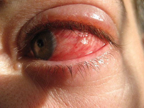
Conjunctivitis is an inflammation or infection of the conjunctiva, the thin transparent layer of tissue that lines the inner surface of the eyelid and covers the white part of the eye. Conjunctivitis, often called “pink eye,” is a common eye disease, especially in children. It may affect one or both eyes. Some forms of conjunctivitis can be highly contagious and easily spread in schools and at home. While conjunctivitis is usually a minor eye infection, sometimes it can develop into a more serious problem.
Conjunctivitis may be caused by a viral or bacterial infection. It can also occur due to an allergic reaction to irritants in the air like pollen and smoke, chlorine in swimming pools, and ingredients in cosmetics or other products that come in contact with the eyes. Sexually transmitted diseases like Chlamydia and gonorrhea are less common causes of conjunctivitis.
People with conjunctivitis may experience the following symptoms:
  • A gritty feeling in one or both eyes
  • Itching or burning sensation in one or both eyes
  • Excessive tearing
  • Discharge coming from one or both eyes
  • Swollen eyelids
  • Pink discoloration to the whites of one or both eyes
  • Increased sensitivity to light
View Image

View Image

What causes conjunctivitis?

The cause of conjunctivitis varies depending on the offending agent. There are three main categories of conjunctivitis: allergic, infectious and chemical:

Allergic Conjunctivitis

  • Allergic Conjunctivitis occurs more commonly among people who already have seasonal allergies. At some point they come into contact with a substance that triggers an allergic reaction in their eyes.
  • Giant Papillary Conjunctivitis is a type of allergic conjunctivitis caused by the chronic presence of a foreign body in the eye. This condition occurs predominantly with people who wear hard or rigid contact lenses, wear soft contact lenses that are not replaced frequently, have an exposed suture on the surface or the eye, or have a glass eye.
Allergic Conjunctivitis occurs more commonly among people who already have seasonal allergies.

Allergic Conjunctivitis occurs more commonly among people who already have seasonal allergies.


Infectious Conjunctivitis

  • Bacterial Conjunctivitis is an infection most often caused by staphylococcal or streptococcal bacteria from your own skin or respiratory system. Infection can also occur by transmittal from insects, physical contact with other people, poor hygiene (touching the eye with unclean hands), or by use of contaminated eye makeup and facial lotions.
  • Viral Conjunctivitis is most commonly caused by contagious viruses associated with the common cold. The primary means of contracting this is through exposure to coughing or sneezing by persons with upper respiratory tract infections. It can also occur as the virus spreads along the body’s own mucous membranes connecting lungs, throat, nose, tear ducts, and conjunctiva.
  • Ophthalmia Neonatorum is a severe form of bacterial conjunctivitis that occurs in newborn babies. This is a serious condition that could lead to permanent eye damage unless it is treated immediately. Ophthalmia neonatorum occurs when an infant is exposed to Chlamydia or gonorrhea while passing through the birth canal. 
View Image


Chemical Conjunctivitis

Chemical Conjunctivitis can be caused by irritants like air pollution, chlorine in swimming pools, and exposure to noxious chemicals.

View Image

How is conjunctivitis diagnosed?

Conjunctivitis can be diagnosed through a comprehensive eye examination. Testing, with special emphasis on evaluation of the conjunctiva and surrounding tissues, may include:
  • Patient history to determine the symptoms the patient is experiencing, when the symptoms began, and the presence of any general health or environmental conditions that may be contributing to the problem.
  • Visual acuity measurements to determine the extent to which vision may be affected.
  • Evaluation of the conjunctiva and external eye tissue using bright light and magnification.
  • Evaluation of the inner structures of the eye to ensure that no other tissues are affected by the condition.
  • Supplemental testing may include taking cultures or smears of conjunctival tissue, particularly in cases of chronic conjunctivitis or when the condition is not responding to treatment.
Using the information obtained from these tests, your optometrist can determine if you have conjunctivitis and advise you on treatment options.

Comprehensive Eye Exam

Conjunctivitis can be diagnosed through a comprehensive eye examination.

How is conjunctivitis treated?

Treatment of conjunctivitis is directed at three main goals:
  1. To increase patient comfort.
  2. To reduce or lessen the course of the infection or inflammation.
  3. To prevent the spread of the infection in contagious forms of conjunctivitis.
The appropriate treatment for conjunctivitis depends on its cause:
  • Allergic conjunctivitis – The first step should be to remove or avoid the irritant, if possible. Cool compresses and artificial tears sometimes relieve discomfort in mild cases. In more severe cases, non-steroidal anti-inflammatory medications and antihistamines may be prescribed. Cases of persistent allergic conjunctivitis may also require topical steroid eye drops.
  • Bacterial conjunctivitis – This type of conjunctivitis is usually treated with antibiotic eye drops or ointments. Improvement can occur after three or four days of treatment, but the entire course of antibiotics needs to be used to prevent recurrence.
  • Viral Conjunctivitis – There are no available drops or ointments to eradicate the virus for this type of conjunctivitis. Antibiotics will not cure a viral infection. Like a common cold, the virus just has to run its course, which may take up to two or three weeks in some cases. The symptoms can often be relieved with cool compresses and artificial tear solutions. For the worst cases, topical steroid drops may be prescribed to reduce the discomfort from inflammation, but do not shorten the course of the infection. Some doctors may perform an ophthalmic iodine eye wash in the office in hopes of shortening the course of the infection. This newer treatment has not been well studied yet, therefore no conclusive evidence of the success exists.
  • Chemical Conjunctivitis – Treatment for chemical conjunctivitis requires careful flushing of the eyes with saline and may require topical steroids. The more acute chemical injuries are medical emergencies, particularly alkali burns, which can lead to severe scarring, intraocular damage or even loss of the eye. 

Contact Lens Wearers

Contact lens wearers may need to discontinue wearing their lenses while the condition is active. Your doctor can advise you on the need for temporary restrictions on contact lens wear.
If the conjunctivitis developed due to wearing contact lenses, your eye doctor may recommend that you switch to a different type of contact lens or disinfection solution. Your optometrist might need to alter your contact lense prescription to a type of lens that you replace more frequently to prevent the conjunctivitis from recurring.

Contact lens wearers may need to discontinue wearing their lenses while the conjunctivitis is active.

Contact lens wearers may need to discontinue wearing their lenses while the conjunctivitis is active.

Self-care

Practicing good hygiene is the best way to control the spread of conjunctivitis. Once an infection has been diagnosed, follow these steps:
  • Don't touch your eyes with your hands.
  • Wash your hands thoroughly and frequently.
  • Change your towel and washcloth daily, and don't share them with others.
  • Discard eye cosmetics, particularly mascara.
  • Don't use anyone else's eye cosmetics or personal eye-care items.
  • Follow your eye doctor's instructions on proper contact lens care.
You can soothe the discomfort of viral or bacterial conjunctivitis by applying warm compresses to your affected eye or eyes. To make a compress, soak a clean cloth in warm water and wring it out before applying it gently to your closed eyelids.
For allergic conjunctivitis, avoid rubbing your eyes. Instead of warm compresses, use cool compresses to soothe your eyes. Over the counter eye drops are available. Antihistamine eye drops should help to alleviate the symptoms, and lubricating eye drops help to rinse the allergen off of the surface of the eye.
See your doctor of optometry when you experience conjunctivitis to help diagnose the cause and the proper course of action.

CoRn & CaLLuS

Corns develop to protect your feet from the hardships they are exposed to.You tend to work day in day out with your feet and hands in mud and otherwise. Your feet get exposed. To protect the feet from dirt, corns and calluses ( thick hardened layers of skins) develop, which if cause discomfort, need to be treated.


View Image


Signs and Symptoms for corn on foot:
You have a corn or callus if you notice:
  • A thick, rough area of skin
  • A hardened,raised bump
  • Tenderness or pain under your skin
  • Flaxy,dry or waxy skin.
Difference between corns and callus.
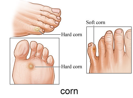
Corns are smaller than calluses and have a hard center surrounded by inflamed skin. They develop on parts of bodythat do not carry any weight, such as tops and sides of your toes. If corns are pushed then they can cause pain or suffer from a dull ache. Calluses usually develop on the soles of the feet, especially under the heels or balls, on the palms, or the knees. They are rarely painful and vary in size and shape. They can be more than inch in diameter, making them larger than corns.
CALLUS
View Image
View Image
CORN
View Image
Causes of Corns and Calluses
  • Ill-fitting shoes: When shoes are too tight, then your feet get compressed causing discomfort and corns and calluses develop. If the shoes are too loose, then the feet rub against the shoe. The foot may rub against a poorly placed seam or stitch inside the shoe.
  • Skipping socks- Wearing shoes or sandals without socks can be a problem. Socks should fit properly.
  • Using hand tools- The use of tools with your hands can also cause calluses on them.
Risk Factors that increase the incidence of calluses and corns on foot:
  • Bunions or hammertoe- A bunion is an abnormal, bony bump that forms on the joint at the base of the big toe. A hammertoe is a toe deformity in which your toe becomes curled up like a claw. These conditions can cause rubbing of the feet in the shoes.
  • Lack of protection for hands- Using hand tools without gloves can cause friction in the hands leading to corns and calluses.
  • Foot Deformities- A pre-existing foot deformity, such as bone spur, may cause problems to your feet.
Treatment of Calluses and Foot Corns:
Severe or persistent corns need to be treated, although they rarely require surgery. Persons suffering from diabetes, or artherosclerosis should not treat a corn on their own, as it can increase the risk of infection. They are acid preparations to reduce corns and calluses. A commonly used preparation is a mixture of salicylic acid,lactic acids and collodion (Duofilm, Salactic Film and Viranol). Over the counter acids are weaker than the ones prescribed.
Acids should not be used by diabetic patients and are not advisable for individuals with impaired sensation or for the elderly with thin skin. Medicated pads are also available. Comfortable shoes should be worn.
Protect your feet from corn and calluses; but if you do suffer then take the necessary precautions to avoid any discomfort. One need not worry about them, if they are not painful.


ASthMA

What is asthma?
Asthma is a chronic, inflammatory disease in which the airways become sensitive to allergens (any substance that triggers an allergic reaction). Several things happen to the airways when a person is exposed to certain triggers:
  • The lining of the airways become swollen and inflamed.
  • The muscles that surround the airways tighten.
  • The production of mucus in increased, leading to mucus plugs.
All of these factors will cause the airways to narrow, thus making it difficult for air to go in and out of your lungs, causing the symptoms of asthma.

What are the symptoms of asthma?

Asthma may resemble other respiratory problems such as emphysema, bronchitis, and lower respiratory infections. It is often under-diagnosed and many people with the disease do not know they have it. Sometimes, the only symptom is a chronic cough, especially at night, or coughing or wheezing that occurs only with exercise. Some people think they have recurrent bronchitis, since respiratory infections usually settle in the chest in a person predisposed to asthma.

What causes asthma?

The exact cause of asthma is not completely known. It is believed to be partially inherited, but it also involves many other environmental, infectious, and chemical factors.
After a person is exposed to a certain trigger, the body releases histamine and other agents that can cause inflammation in the airways. The body also releases other factors that can cause the muscles of the airways to tighten, or become smaller. There is also an increase in mucus production that may clog the airways.
Some persons have exercise-induced asthma, which is caused by varying degrees of exercise. Symptoms can occur during, or shortly after, exercise. Each person has different triggers that cause the asthma to worsen. You should discuss this with your physician.
The changes that occur in asthma are believed to happen in two phases:
  1. An immediate response to the trigger leads to swelling and narrowing of the airways. This makes it initially difficult to breathe.
  2. A later response, which can happen four to eight hours after the initial exposure to the allergen, leads to further inflammation of the airways and obstruction of airflow.
Knowing your risk factors to any disease can help to guide you into the appropriate actions, including changing behaviors and being clinically monitored for the disease.

What are the risk factors for an asthma attack?

Although anyone may have an asthma attack, it most commonly occurs in the following persons:
  • children by the age of 5
  • adults in their 30s
  • adults older than 65
  • people living in urban communities
Other factors include the following:
  • family history of asthma
  • personal medical history of allergies
Children most susceptible to asthma attacks include the following:
  • children with a family history of asthma
  • children who have allergies
  • children who have exposure to secondhand tobacco smoke

What happens during an asthma attack or asthma exacerbation?

Persons with asthma have acute episodes when the air passages in their lungs become narrower, and breathing becomes more difficult. These problems are caused by an over-sensitivity of the lungs and airways.
  • The lungs and airways overreact to certain triggers causing:
    • the lining of the airways to become inflamed and swollen.
    • tightening of the muscles that surround the airways.
    • an increased production of mucus.
  • Breathing becomes harder and may hurt.
  • There may be coughing.
  • There may be a wheezing or whistling sound, which is typical of asthma. Wheezing occurs because of the rush of air which moves through the narrowed airways.

How is asthma diagnosed?

To diagnose asthma and distinguish it from other lung disorders, physicians rely on a combination of medical history, physical examination, and laboratory tests, which may include the following:
  • spirometry - a spirometer is a device used by your physician that assesses lung function. Spirometry, the evaluation of lung function with a spirometer, is one of the simplest, most common pulmonary function tests and may be necessary for any/all of the following reasons:

    • to determine how well the lungs receive, hold, and utilize air
    • to monitor a lung disease
    • to monitor the effectiveness of treatment
    • to determine the severity of a lung disease
    • to determine whether the lung disease is restrictive (decreased airflow) or obstructive (disruption of airflow)
  • peak flow monitoring (PFM) - a device used to measure the fastest speed in which a person can blow air out of the lungs. During an asthma or other respiratory flare up, the large airways in the lungs slowly begin to narrow. This will slow the speed of air leaving the lungs and can be measured by a PFM. This measurement is very important in evaluating how well or how poorly the disease is being controlled.
  • chest x-rays - a diagnostic test which uses invisible electromagnetic energy beams to produce images of internal tissues, bones, and organs onto film.
  • blood tests - to analyze the amount of carbon dioxide and oxygen in the blood.
  • allergy tests

What are the triggers that can cause an asthma attack?

According to the National Institute for Allergy and Infectious Diseases, the American Academy of Allergy, Asthma, and Immunology, and other organizations, triggers for asthma include the following:
Allergens
  • pollen
  • mold
  • animal protein (dander, urine, oil from skin)
  • house dust/dust mites
  • cockroaches
  • certain foods
Respiratory Infections and Sinusitis
Infections can cause irritation of the airways, nose, throat, lungs, and sinuses, and worsens asthma.
Irritants
  • strong odors and sprays, such as perfumes, household cleaners, cooking fumes, paints, and varnishes
  • chemicals such as coal, chalk dust, or talcum powder
  • air pollutants
  • changing weather conditions, including changes in temperature, barometric pressure, humidity, and strong winds
  • Chemical-exposure on the job, such as occupational vapors, dust, gases, or fumes.
Sensitivity to Medications
Medications, such as aspirin and sulfites, cause up to 20 percent of adult asthmatic attacks as a result of sensitivities or allergies to them. These medications often include:
  • aspirin
  • other non-steroidal anti-inflammatory medications, such as ibuprofen, indomethacin, naproxen
  • sulfites used as preservatives in food and beverage
Before taking any medication, including over-the-counter medications, consult your physician.
Exercise
Exercise can trigger an asthma attack, often because of the inhaled cool and dry air. Long-term strenuous activities such as long distance running, are most likely to induce asthma, and swimming is the least likely.
Gastroesophageal reflux
GERD, a condition characterized by persistent reflux of stomach acids, is common in individuals with asthma. Symptoms may include heartburn, belching, or spitting up in infants.
Smoke
Tobacco smoke, whether directly or passively inhaled, has been shown to worsen asthma.
Wood smoke from wood-burning heating stoves and fireplaces can release irritating chemicals such as sulfur dioxide.
Emotional Anxiety and Nervous Stress
Reactions from stress and anxiety are considered to be more of an effect than a cause. They can cause fatigue, which may affect the immune system and, in turn, increase either asthma symptoms or bring on an attack.


Go to fullsize image    


  View Image

Go to fullsize image

Tuesday, August 17, 2010

bReasT canCer...


WHAT IS THE BREAST CANCER????


Breast cancer is a kind of tumor that develops in the cells of a person's breast. You may think that only women can get breast cancer, but because all people have breast tissue, men can get breast cancer as well - though this is very rare.


RISK FACTOR OF BREAST CANCER

Gender

-> Simply being a woman is the main risk factor for developing breast cancer. Although women have many more breast cells than men, the main reason they develop more breast cancer is because their breast cells are constantly exposed to the growth-promoting effects of the female hormones estrogen and progesterone.

Aging

->  Your risk of developing breast cancer increases as you get older. About 1 out of 8 invasive breast cancers are found in women younger than 45, while about 2 out of 3 invasive breast cancers are found in women age 55 or older.

Menstrual periods

->  Women who have had more menstrual cycles because they started menstruating at an early age (before age 12) and/or went through menopause at a later age (after age 55) have a slightly higher risk of breast cancer. This may be related to a higher lifetime exposure to the hormones estrogen and progesterone.

Not having children, or having them later in life

->  Women who have had no children or who had their first child after age 30 have a slightly higher breast cancer risk. Having many pregnancies and becoming pregnant at a young age reduce breast cancer risk. Pregnancy reduces a woman's total number of lifetime menstrual cycles, which may be the reason for this effect.

Recent oral contraceptive use

->  Studies have found that women using oral contraceptives (birth control pills) have a slightly greater risk of breast cancer than women who have never used them. This risk seems to decline back to normal over time once the pills are stopped. Women who stopped using oral contraceptives more than 10 years ago do not appear to have any increased breast cancer risk. 

Not breast-feeding

->  Some studies suggest that breast-feeding may slightly lower breast cancer risk, especially if breast-feeding is continued for 1½ to 2 years. But this has been a difficult area to study, especially in countries such as the United States, where breast-feeding for this long is uncommon

Alcohol

->  Use of alcohol is clearly linked to an increased risk of developing breast cancer. The risk increases with the amount of alcohol consumed. Compared with non-drinkers, women who consume 1 alcoholic drink a day have a very small increase in risk

Being overweight or obese

->  Being overweight or obese has been found to increase breast cancer risk, especially for women after menopause. Before menopause your ovaries produce most of your estrogen, and fat tissue produces a small amount of estrogen. After menopause (when the ovaries stop making estrogen), most of a woman's estrogen comes from fat tissue. Having more fat tissue after menopause can increase your chance of getting breast cancer by raising estrogen levels.

Lack of physical activity

->  Evidence is growing that physical activity in the form of exercise reduces breast cancer risk. The main question is how much exercise is needed. In one study from the Women's Health Initiative (WHI) as little as 1.25 to 2.5 hours per week of brisk walking reduced a woman's risk by 18%. Walking 10 hours a week reduced the risk a little more.

High-fat diets

->  More research is needed to better understand the effect of the types of fat eaten on breast cancer risk. But it is clear that calories do count, and fat is a major source of these. High-fat diets can lead to being overweight or obese, which is a breast cancer risk factor. A diet high in fat has also been shown to influence the risk of developing several other types of cancer, and intake of certain types of fat is clearly related to heart disease risk.

Bras

->  Internet e-mail rumors and at least one book have suggested that bras cause breast cancer by obstructing lymph flow. There is no good scientific or clinical basis for this claim. Women who do not wear bras regularly are more likely to be thinner, which would probably contribute to any perceived difference in risk.

Tobacco smoke

->  An active focus of research is whether secondhand smoke increases the risk of breast cancer. Both mainstream and secondhand smoke contain chemicals that, in high concentrations, cause breast cancer in rodents. Chemicals in tobacco smoke reach breast tissue and are found in breast milk.

Night work

->  Several studies have suggested that women who work at night -- for example, nurses on a night shift -- may have an increased risk of developing breast cancer. This is a fairly recent finding, and more studies are looking at this issue. Some researchers think the effect may be due to changes in levels of melatonin, a hormone whose production is affected by the body's exposure to light, but other hormones are also being studied.

                                                  *******************************

As prevention, we must do Breast Self Examination (BSE) at least every year for women 40 years old and above and every 3years for women 20 until 30 years old.


HOW TO DO BREAST SELF EXAMINATION???

  • Lie down and place your right arm behind your head. The exam is done while lying down, not standing up. This is because when lying down the breast tissue spreads evenly over the chest wall and is as thin as possible, making it much easier to feel all the breast tissue.
  • Use the finger pads of the 3 middle fingers on your left hand to feel for lumps in the right breast. Use overlapping dime-sized circular motions of the finger pads to feel the breast tissue.
 
  • Use 3 different levels of pressure to feel all the breast tissue. Light pressure is needed to feel the tissue closest to the skin; medium pressure to feel a little deeper; and firm pressure to feel the tissue closest to the chest and ribs. It is normal to feel a firm ridge in the lower curve of each breast, but you should tell your doctor if you feel anything else out of the ordinary. If you're not sure how hard to press, talk with your doctor or nurse. Use each pressure level to feel the breast tissue before moving on to the next spot.
  • Move around the breast in an up and down pattern starting at an imaginary line drawn straight down your side from the underarm and moving across the breast to the middle of the chest bone (sternum or breastbone). Be sure to check the entire breast area going down until you feel only ribs and up to the neck or collar bone (clavicle).
  • There is some evidence to suggest that the up-and-down pattern (sometimes called the vertical pattern) is the most effective pattern for covering the entire breast, without missing any breast tissue.
  • Repeat the exam on your left breast, putting your left arm behind your head and using the finger pads of your right hand to do the exam.
  • While standing in front of a mirror with your hands pressing firmly down on your hips, look at your breasts for any changes of size, shape, contour, or dimpling, or redness or scaliness of the nipple or breast skin. (The pressing down on the hips position contracts the chest wall muscles and enhances any breast changes.)
  • Examine each underarm while sitting up or standing and with your arm only slightly raised so you can easily feel in this area. Raising your arm straight up tightens the tissue in this area and makes it harder to examine